Abstract
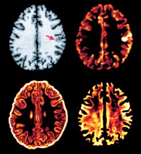
Focal cortical dysplasia (FCD), a malformation of cortical development, is a frequent cause of pharmacologically intractable epilepsy. FCD is characterized on Tl-weighted MRI by cortical thickening, blurring of the gray-matter/white-matter interface, and gray-level hyperintensity. We have previously used computational models of these characteristics to enhance visual lesion detection. In the present study we seek to improve our methods by combining these models with features derived from texture analysis of MRI, which allows measurement of image properties not readily accessible by visual analysis. These computational models and texture features were used to develop a two-stage Bayesian classifier to perform automated FCD lesion detection. Eighteen patients with histologically confirmed FCD and 14 normal controls were studied. On the MRI volumes of the 18 patients, 20 FCD lesions were manually labeled by an expert observer. Three-dimensional maps of the computational models and texture features were constructed for all subjects. A Bayesian classifier was trained on the computational models to classify voxels as cerebrospinal fluid, gray-matter, white-matter, transitional, or lesional. Voxels classified as lesional were subsequently reclassified based on the texture features. This process produced a 3D lesion map, which was compared to the manual lesion labels. The automated classifier identified 17/20 manually labeled lesions. No lesions were identified in controls. Thus, combining models of the T1-weighted MRI characteristics of FCD with texture analysis enabled successful construction of a classifier. This computer-based, automated method may be useful in the presurgical evaluation of patients with severe epilepsy related to FCD.

