Abstract
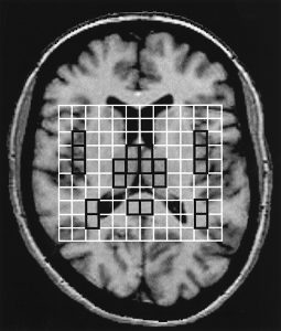
Experimental work in animal models of generalized epilepsy and clinical data in humans with idiopathic generalized epilepsy (IGE) indicate that the thalamo-cortical circuitry is involved in the generation of epileptic activity. The purpose of this study was to evaluate in vivo the chemical and structural integrity of the thalamus in patients with IGE. Thalamic proton magnetic resonance spectroscopic imaging (1H-MRSI), measuring N-acetylaspartate (NAA), choline-containing compounds and creatine (Cr) was performed in 20 IGE patients and in a group of age-matched healthy subjects. Additionally, 1H-MRSI measurements were taken in the insular cortex, the posterior temporal lobe white matter and the splenium of the corpus callosum. MRI volumetric analysis of the thalamus was performed in all patients. At the time of the examination, seizures were well controlled in 10 IGE patients and poorly controlled in nine. One patient was newly diagnosed and had the MRI and MRSI examination prior to starting the antiepileptic medication. In IGE patients, 1H-MRSI showed a reduction of mean thalamic NAA/Cr compared with normal controls; no difference was found in NAA/Cr in the other examined areas. There was no difference in NAA/Cr between patients whose seizures were well controlled and those in whom seizures were not controlled. There was no correlation between thalamic NAA/Cr and mean number of spike and wave complexes. We found a significant negative correlation between thalamic NAA/Cr and duration of epilepsy. The mean thalamic volume in patients with IGE was not different from normal controls. These results show evidence of progressive thalamic neuronal dysfunction in patients with IGE supporting the notion of abnormal thalamo-cortical circuitry as a substrate of seizure generation in this form of epilepsy. The thalamic dysfunction may occur regardless of amount of spike and wave activity.

